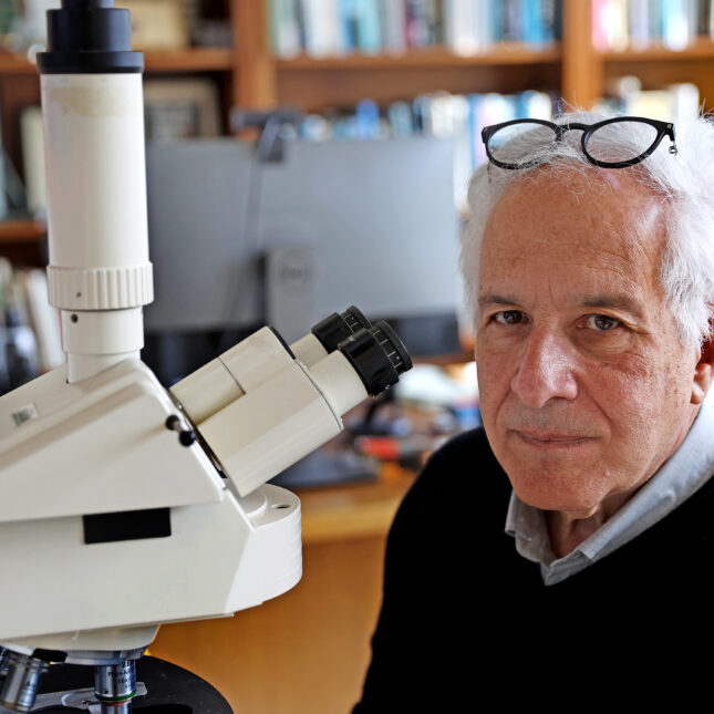
About 10 years ago, a small piece of human brain arrived in the lab of Dr. Jeffrey Lichtman at Harvard. It came directly from an operating room of a nearby hospital, where it was excised from an epilepsy patient undergoing a procedure to reduce her seizures.
In the years that followed, Lichtman’s team methodically reconstructed the byzantine wiring patterns of the brain by feeding the 1-cubic-millimeter sample into a $6 million device that sliced it into impossibly-thin slivers. Then, using images of those slivers taken by electron microscopy, they painstakingly recreated the intricate latticework connecting individual cells to one another.
The result is the most-detailed digital map, or “connectome,” of the human brain ever created.

This article is exclusive to STAT+ subscribers
Unlock this article — plus in-depth analysis, newsletters, premium events, and networking platform access.
Already have an account? Log in
Already have an account? Log in
To submit a correction request, please visit our Contact Us page.


STAT encourages you to share your voice. We welcome your commentary, criticism, and expertise on our subscriber-only platform, STAT+ Connect